-
Welcome back Guest! Did you know you can mentor other members here at H-M? If not, please check out our Relaunch of Hobby Machinist Mentoring Program!
- Forums
- THE PROJECTS AREA
- PROJECT OF THE DAY --- WHAT DID YOU DO IN YOUR SHOP TODAY?
- Project of the Day Mega-Thread Archives
You are using an out of date browser. It may not display this or other websites correctly.
You should upgrade or use an alternative browser.
You should upgrade or use an alternative browser.
2018 POTD Thread Archive
- Thread starter 2volts
- Start date
- Joined
- Nov 23, 2014
- Messages
- 2,634
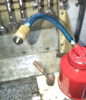
Hi Savarin,Had to make it straight away, brilliant, such a simple concept.
View attachment 283157
Nice job! Only thing I did differently was to cut away half of the tip. It gives the oil a place to go to get around the ball. A needle file V-notch on the tip would do the same thing. The concept is really stolen from a shop vac nozzle. Ours have a relief notch on the side of the opening so it doesn't seal to the floor.
Bruce
- Joined
- Aug 22, 2012
- Messages
- 4,271
Hi Bruce,
I was in such a hurry to test it I never got that far but I did make sure the tip is short enough so it doesnt set tight up against the ball and the oil pressure would push the ball out the way, It does and I am wrapped with the simplicity of the whole project.
Why dont they come with this from the factory.
- Joined
- Apr 23, 2018
- Messages
- 6,873
Hi Pontiac428,
Bits look great. Which cutter/grinder did you pick up from Shars?
Thanks, Bruce
Bruce, It's their Deckel clone. It comes with work heads and fixtures that expand the little grinder's functionality. It's not German craftsmanship, but I'm excited to encounter those "I can do that" moments that happen when you have the right equipment!
(from mobile)
- Joined
- Sep 28, 2013
- Messages
- 4,395
made the wife's Christmas present, a mortar and pestle. She had a small one years ago but it disappeared in one of our many moves and she's missed it, so I figured that this would make a nice surprise for her.
Making the mortar was a bloody miserable job. Used the trusty rusty 4x6 to cut 3 1/4" off a chunk of 3" supposedly stainless round (that's what the scrapyard said and charged me for, but it's magnetic), then step drilled the hole out to 1" on the mill. Faced off the bottom and turned it round in the lathe to start boring out the cavity. Several days later, ta da!

next to parent material

used math (well, technically a Math professor) in Excel to calculate all the bore depths and radii for the hemisphere at the bottom in 2.5 thou increments. Didn't exactly go to plan as you can see but the "texture" should help with the grinding, right?
donor material for the pestle. Definitely stainless and they periodically show up at the scrap yard. not sure what it's for, but it's well machined and looks like there's the remnant of a carbide tip on the end. Who knows? Anyway, the future pestle is the piece in the middle

turned the business end with my boring head ball turner

then intentionally added some grooves for extra grindingness

first time ever using the cup center to turn the taper and tidy up the flank of the pestle

all done! The hand end of the pestle didn't come out quite the way I intended, but it'll do


it's now wrapped up and stopping the Christmas tree from blowing away
Merry Christmas!
Making the mortar was a bloody miserable job. Used the trusty rusty 4x6 to cut 3 1/4" off a chunk of 3" supposedly stainless round (that's what the scrapyard said and charged me for, but it's magnetic), then step drilled the hole out to 1" on the mill. Faced off the bottom and turned it round in the lathe to start boring out the cavity. Several days later, ta da!
next to parent material
used math (well, technically a Math professor) in Excel to calculate all the bore depths and radii for the hemisphere at the bottom in 2.5 thou increments. Didn't exactly go to plan as you can see but the "texture" should help with the grinding, right?
donor material for the pestle. Definitely stainless and they periodically show up at the scrap yard. not sure what it's for, but it's well machined and looks like there's the remnant of a carbide tip on the end. Who knows? Anyway, the future pestle is the piece in the middle
turned the business end with my boring head ball turner
then intentionally added some grooves for extra grindingness
first time ever using the cup center to turn the taper and tidy up the flank of the pestle
all done! The hand end of the pestle didn't come out quite the way I intended, but it'll do
it's now wrapped up and stopping the Christmas tree from blowing away
Merry Christmas!
In the shop a bit this morning. I’ve been working on repurposing a medication bottle. I take some maintenance meds and their mailed to me. This small no spill oil container is for my bench for hand tapping

Sent from my iPhone using Tapatalk

Sent from my iPhone using Tapatalk
- Joined
- Sep 28, 2013
- Messages
- 4,395
Also got a couple of other projects done. One was adapting a flask holder to an orbital shaker for the lab, so we can use one of our incubators to grow bacterial cultures. Both parts were crazy cheap off Amazon, so it was worth the effort.


Second was spending 3 days at the beginning of the week with a very nice guy from Olympus to test drive a laser scanning confocal microscope that I'm rewriting a grant for. He brought an all singing all dancing version with him that is about $550,000, but we're asking for a simpler one for just $330,000
here's a mouse brain lesion. Blue = nuclei, green = actin (a cytoskeleton filament), not sure what red is labelling (colleagues work)

next up is an autofluorescence image of a fire worm (polychaete annelid) - the spikes are calciferous and break off in your skin causing either burning itching sensations or long term numbness

close up of the spikes (chitae?). My colleage was super excited by this image as some of the autofluorescence is seen only in the tips of the spikes, which might be the causative agent of the numbness. Long way down the road, but that might lead to new anaesthetics. Or not, given that it took over 18 months for the numbness in her finger tips to wear off when she was collecting them.

now my stuff:
a green fluorescent protein expressed in neurons (if you look closely you can see axons and dendrites)

a different construct expressed in the germ cell niches at the end of the worm gonads

and a final one expressed in the intestinal cells - the nuclei are the bright green circles

I love playing with microscopes, you can get some truly beautiful images
Second was spending 3 days at the beginning of the week with a very nice guy from Olympus to test drive a laser scanning confocal microscope that I'm rewriting a grant for. He brought an all singing all dancing version with him that is about $550,000, but we're asking for a simpler one for just $330,000
here's a mouse brain lesion. Blue = nuclei, green = actin (a cytoskeleton filament), not sure what red is labelling (colleagues work)
next up is an autofluorescence image of a fire worm (polychaete annelid) - the spikes are calciferous and break off in your skin causing either burning itching sensations or long term numbness
close up of the spikes (chitae?). My colleage was super excited by this image as some of the autofluorescence is seen only in the tips of the spikes, which might be the causative agent of the numbness. Long way down the road, but that might lead to new anaesthetics. Or not, given that it took over 18 months for the numbness in her finger tips to wear off when she was collecting them.
now my stuff:
a green fluorescent protein expressed in neurons (if you look closely you can see axons and dendrites)
a different construct expressed in the germ cell niches at the end of the worm gonads
and a final one expressed in the intestinal cells - the nuclei are the bright green circles
I love playing with microscopes, you can get some truly beautiful images
Last edited:
- Joined
- May 2, 2018
- Messages
- 152
- Joined
- Jul 14, 2017
- Messages
- 2,455
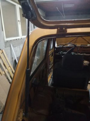
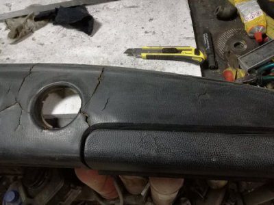
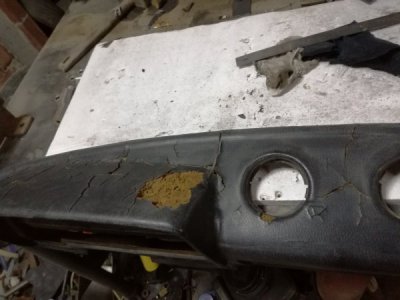
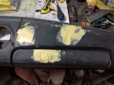
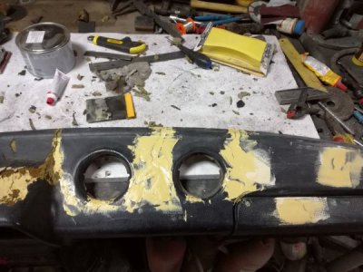
Last couple of days i've been working on the little Lada, working on smaller jobs first of which was mounting the trunk lid and drilling, tapping and installing gas struts, i had to modify the mounting holes to accept the new style ball mounts, this left me with a empty workbench, so i got the dashboard on there to work on fixing it, i've had no luck in finding a new dashboard so i'm using body filler to fill the holes and plan to use bedliner to apply a textured coat over it. I may need to start more than one job at a time because the body filler seams to take long time to harden, probably because of the cold.
